Multi Model treatment
A Case Study
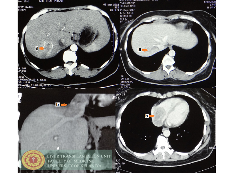
A 42 year old diabetic lady presented with recent history of ankle swelling and shortness of breath. On further evaluation she was found to have Child's class A cirrhosis with a model for end stage liver disease score of 9. Liver imaging and echocardiogram showed a typical hepatocellular carcinoma in segment VIII of the liver extending in to the right hepatic vein, inferior vena cava (IVC) and the right atrium (figure 1). There was a secondary thrombus in the IVC. Right atrial inflow and outflow showed features of impending obstruction. Considering her excellent general condition, it was decided to perform combine thoraco-laparotomy.
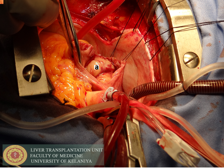
During laparotomy liver had significant macroscopic changes of cirrhosis. It was decided to abandon the liver resection and to remove atrial tumor to palliate symptoms. The right atrium was opened under cardiopulmonary bypass and the atrial tumor was removed from its origin at right hepatic vein (figure 2). Secondary clot in the IVC was also removed.
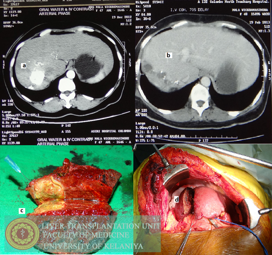
Immediately, on the 5th post-operative day she underwent trans-arterial chemo embolization of the tumor. This was followed by a second TACE session after six weeks (figure 3). In the follow-up CT after 3 months, right portal vein appeared thrombosed. The right lobe was atrophic and there was significant hypertrophy of the left lobe (figure 3). Based on these findings, she subsequently underwent right hepatectomy. She is recurrence free at 20 months after the initial surgery.
Tumors with major vascular invasion are considered contraindication for surgery (1). Invasion of left atrium is an extreme situation. Overall survival in them is reported to be less than three months (2). Only few cases are reported to be successfully resected. Approach in this patient was unique. Given the patients clinical situation, we had to manage the patient in stages. This case highlights that decisions are best taken on individual basis. Combination of one treatment modality with another is becoming popular (3). Multimodal approach gave us the chance to control the primary tumor and offer curative resection in a later stage.
References
- Forner A, Reig ME, de Lope CR, Bruix J. Current strategy for staging and treatment: the BCLC update and future prospects. Semin Liver Dis. 2010;30:61–74
- Kim SU, Kim YR, Kim do Y, Kim JK, Lee HW, Kim BK, et.al. Clinical features and treatment outcome of advanced hepatocellular carcinoma with inferior vena caval invasion or atrial tumor thrombus. Korean J Hepatol. 2007 Sep;13(3):387-95.
- Cabibbo G, Latteri F, Antonucci M, Craxì A. Multimodal approaches to the treatment of hepatocellular carcinoma.Nat ClinPract Gastroenterol Hepatol. 2009 Mar;6(3):159-69
Captions

- Tumor in segment VIII. Arterial phase before surgery
- Tumor invading the right hepatic vein,
- Tumor extension in to the inferior vena cava,
- Tumor in the left atrium

Intra operative picture of the tumor
- in the opened left atrium

- Tumor after first trans arterial chemoembolisation (TACE) also showing left lobe hypertrophy,
- Computed tomogram after right hepatectomy,
- Right hepatectomy specimen after two cycles of TACE, tumor had 100% necrosis under microscopy,
- Intra operative view after splitting the liver.
